When Your Child Has an Atrioventricular (AV) Canal Defect
The heart is divided into 4 chambers. The 2 upper chambers are called atria and the 2 lower chambers are called ventricles. The heart contains 4 valves. The valves open and close to keep blood flowing forward through the heart. An atrioventricular (AV) canal defect is a large hole in the center of the heart. It’s caused by the following problems with heart structure:
-
Atrial septal defect (ASD). A hole in the dividing wall (atrial septum) that separates the atria in the heart.
-
Ventricular septal defect (VSD). A hole in the dividing wall (ventricular septum) that separates the ventricles in the heart.
-
A single atrioventricular (AV) valve. A single valve that develops in place of the separate tricuspid (right side) and mitral valves (left side). These valves control the flow of blood from the atria to the ventricles.
When all of these heart problems are present, the AV canal defect is considered complete. The healthcare provider can tell you more about your child’s heart and the exact defects they have. An AV canal defect can usually be treated.
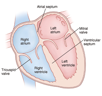 |
| Normal heart. |
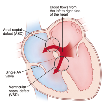 |
| An AV canal defect causes more blood than normal to pass through the right side of the heart and lungs through an ASD and VSD. |
What causes an atrioventricular canal defect?
An AV canal defect is a congenital heart defect. This means it is a problem with the heart’s structure that your child was born with. The exact cause is unknown. Children with Down syndrome (trisomy 21) are at higher risk of having this heart defect.
Why is an atrioventricular canal defect a problem?
Blood normally flows from chamber to chamber in one direction through the left and right sides of the heart. With an AV canal defect, blood flows through the ASD and VSD from the left side of the heart to the right side. This is called a left-to-right shunt. It causes more blood than normal to pass through the right side of the heart. It causes the left side of the heart to become enlarged (dilated). More blood than normal has to be pumped to the lungs. Over time, the extra blood flow causes the lungs to become filled with extra blood and fluid. This leads to a condition called congestive heart failure (CHF). Problems can also occur with the single AV valve. Because it's malformed, blood may leak backward from the ventricles to the atria (valve insufficiency or regurgitation). This causes the heart to work even harder.
What are the symptoms?
Children with an AV canal defect usually have signs and symptoms of heart failure in infancy. These include:
-
Tiredness
-
Trouble breathing or rapid breathing
-
Trouble feeding
-
Poor weight gain and growth
-
Sweating
-
Blue or purple coloring of the lips, skin, and nails (cyanosis)
-
Heart murmur
Depending on how serious the defect is and several other factors, symptoms may not show up until adulthood.
How is an atrioventricular canal defect diagnosed?
Typically, a complete AV canal defect can be diagnosed in a fetus with a fetal echocardiogram. This can be performed as early as 12 weeks gestation. If the AV canal defect is not diagnosed in the fetus, the healthcare provider checks for signs of a heart problem, such as a heart murmur during a physical exam. This is an extra noise caused when blood doesn’t flow smoothly through the heart. If a heart problem is suspected, your child may be referred to a pediatric cardiologist. This is a doctor with special training to diagnose and treat heart problems in children. To check for an AV canal defect, the following tests may be done:
-
Chest X-ray. X-rays are used to take a picture of the heart and lungs.
-
Electrocardiogram (ECG). The test records the electrical activity of the heart.
-
Echocardiogram (echo). Sound waves (ultrasound) are used to create a picture of the heart and look for structural defects.
How is an atrioventricular canal defect treated?
Children with a complete AV canal defect are treated early in infancy with heart surgery, usually within the first 6 months of life. Medicines may be prescribed to help manage symptoms until surgery is scheduled. These can include:
-
Water pills (diuretics). This helps rid the body of excess water. This reduces fluid in the lungs and may improve breathing.
-
Digoxin. It helps the heart pump blood with more force. This improves how the heart works.
-
ACE inhibitors. This makes blood vessels relax and allows blood to flow more easily from the heart.
-
Nutrition. A baby with an atrioventricular canal defect may need supplemental nutrition with a feeding tube.
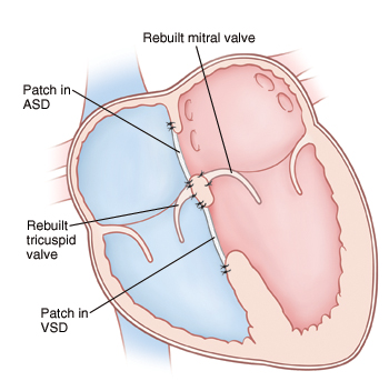 |
| The ASD and VSD are closed with patches. The single AV valve is divided and used to rebuild separate tricuspid and mitral valves. |
Your child’s experience: heart surgery
Heart surgery to repair an AV canal defect is done by a pediatric heart surgeon. The surgery lasts at least 4 to 6 hours. It takes place in an operating room in a hospital. You’ll stay in the waiting room during your child’s surgery:
-
Before surgery. You’ll be told to keep your child from eating or drinking anything for a certain amount of time before surgery. Follow these instructions carefully.
-
During surgery. Your child is given medicine (sedative and anesthesia) to sleep and not feel pain during surgery. A breathing tube is placed in your child’s windpipe (trachea) during this time. Special equipment keeps track of your child’s heart rate, blood pressure, and oxygen levels. Your child is also placed on a heart-lung bypass machine. This allows blood to continue flowing to the body while the heart is stopped so that it can be operated on. An incision is made in the chest through the breastbone (sternum) to access the heart. The ASD and VSD are closed with 1 or 2 patches. The single AV valve is rebuilt into 2 separate valves. Once the repair is complete, your child is taken off the bypass machine and the chest is closed.
-
After the surgery. Your child is taken to a critical care unit to be cared for and watched. Several catheters, tubes, and wires may be attached to your child to assist the medical team in caring for your child. You can stay with your child during this time. They may stay in the hospital for 7 to 10 days. When your child is ready to leave the hospital, you’ll be given directions for home care and follow up.
Risks and complications of heart surgery
-
Reaction to sedative or anesthesia
-
Leakage of blood through one or both of the new valves back into the atria (valve insufficiency); which may need additional surgery.
-
Stenosis of one or both of the new valves, which may need additional surgery.
-
Incomplete closure of the ASD, VSD, or both. This may need more treatment.
-
Abnormal heart rhythm (arrhythmia)
-
Infection
-
Bleeding
-
Nervous system problems
-
Abnormal buildup of fluid around the heart and lungs
-
Death
When to call the healthcare provider
After heart surgery, call the healthcare provider right away if any of the following occur:
-
Increased pain, swelling, redness, bleeding, or drainage of an incision site
-
A fever 100.4°F (38°C), or as directed by your healthcare provider
-
Increased tiredness
-
Fast or irregular breathing
-
Nausea or vomiting that continues
-
A cough that won’t go away
Call 911
Call 911 if any of the following occur:
What are the long-term concerns?
-
After treatment, most children with an AV canal defect can be active. The level and extent of physical activity will vary with each child. Some children may not be able to play certain contact sports, such as football. Check with the cardiologist about what activities are appropriate and safe for your child.
-
Regular follow-up visits with the cardiologist are needed for the rest of your child’s life. The rebuilt valves will always be abnormal. They need to be checked to make sure they’re working correctly without too much leakage, especially the mitral valve. Further valve treatment or replacement may be needed later in life.
-
Depending on the details of the surgical repair, your child may need to take antibiotics before having any surgery or dental work to prevent infection of the inside lining of the heart and valves. This infection is called infective endocarditis. Antibiotics should be taken as directed by the cardiologist.
-
Most children born with AV canal defects today go on to lead productive lives as adults.