Pericardial Window
A pericardial window is a surgery done on the sac around the heart. A small part of the sac is removed. This lets extra fluid drain from the sac.
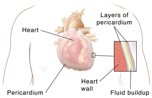
Understanding the sac around the heart
A fibrous sac called the pericardium surrounds the heart. This sac has 2 thin layers with a small amount of fluid in between them. The fluid helps reduce friction between the 2 layers when the heart beats. In some cases, too much fluid builds up between the layers. The extra fluid puts pressure on the heart, making it hard for the heart to pump normally. This can cause dizziness, nausea, and low blood pressure. It can also cause chest pain and trouble breathing. The extra fluid needs to be drained.
Sometimes it’s not clear why the fluid builds up. But the fluid can build up if you have any of these:
-
Infection of the heart or pericardial sac
-
Inflammation of the pericardial sac due to a heart attack
-
Cancer
-
Injury
-
Open heart surgery
-
Immune system disease
-
Reactions to certain medicines
-
Exposure to radiation
-
Kidney failure with uremia
Why a pericardial window is done
Many conditions can cause fluid to build up around the heart. Sometimes it can be treated with medicine. In other cases, the extra fluid is dangerous and needs draining right away.
A pericardial window is one way to remove fluid around the heart. It can:
-
Drain the extra fluid around the heart
-
Prevent fluid from building up too much in the future
-
Let the healthcare provider take a sample of tissue from the sac (biopsy) and diagnose the cause of the extra fluid
Another way to remove fluid is catheter pericardiocentesis. This procedure uses a needle and a long, thin tube (catheter) to drain the fluid from the heart. But some health conditions make this method difficult. Also, extra fluid can come back.
How a pericardial window is done
A pericardial window can be done in a number of ways. In most cases, it’s done under general anesthesia. This means you are in a state like deep sleep during the surgery. A cut (incision) is made under the bottom of the breastbone or between the ribs to get to the pericardium. Sometimes the healthcare provider will make several small incisions on the side of the chest instead. This is called video-assisted thoracoscopy (VATS). VATS uses a small camera and small tools to create the pericardial window through these small holes. Once the cuts are made, the pericardium is opened, the fluid is drained, and a portion of the pericardium is removed. If the procedure is done through the chest, tubes may be placed in the chest after the pericardium is removed to continue draining fluid. These tubes are removed later.
Risks of a pericardial window
All procedures have some risks. The risks of a pericardial window include:
-
Bleeding
-
Infection
-
Blood clot that can lead to stroke or other problems
-
Abnormal heart rhythms that can cause death in rare cases
-
Heart attack
-
Problems from anesthesia
-
Return of the extra fluid
-
Need for a repeat procedure
-
Need for the whole pericardium to be removed
-
Damage to the heart itself
Your own risks may vary according to your age, health, the type of surgery you have, and other factors. Talk with your healthcare provider to find out what risks may apply to you.